Dog Leg Bones Diagram - Dog Leg Anatomy Explained Injury Types And Treatments Medrego Guide / Skull the skeleton of the head not to be confused with:. A small boat propelled by a scull or a pair of sculls. Just as the human muscular system is composed of units of tissue connected to the skeletal system, skin, and other muscles, a dog's muscle anatomy is arranged in a similar fashion. In diagram a, a human man is shown next to a dog, the bones are highlighted on each animal and they are shown to be the same bone but in different proportions and ratios. One extremely important part of a dog's skeletal anatomy is the skull. The tibia and fibula are in the lower leg, between the knee and the foot.
Click on the image for a larger look at it. They are hard, rigid, and are made up of calcium and phosphorous. For ease of reference, anatomists separate these into two divisions: The axial skeleton, which contains the bones along the long axis of the body (i.e., the head and the torso) and the appendicular skeleton, which includes the bones of the appendages. The larger image will open in a new window, use the close button when finished.
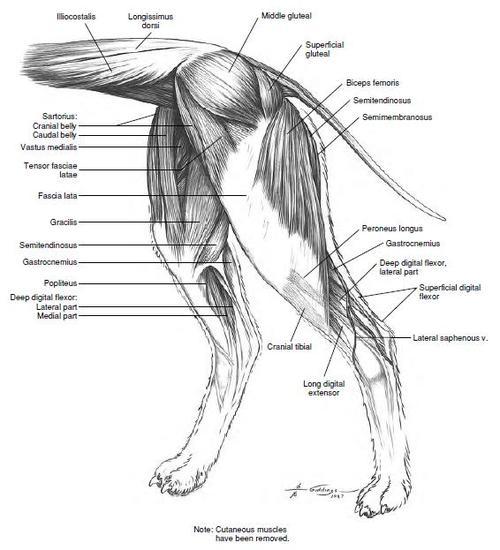
Dog Leg Bones Diagram Dog Muscle Anatomy By Thedragonofdoom On Deviantart What Does This Suggest About Mammals from lh3.googleusercontent.com 172 of the 206 human bones are part of a pair, including all 126 bones of the appendicular skeleton and. The muscular anatomy of a dog, while serving the same purpose in a dog, differs in structure and function from the muscular system in a human body. As with all living beings, the bones surround and protect the internal organs of the body from injury. A small boat propelled by a scull or a pair of sculls. Skull the skeleton of the head not to be confused with: At the knee joint, the femur connects to the tibiotarsus (shin) and fibula (side of lower leg). Innominate bones are evolutionary significant in that they allow birds to lay eggs. One extremely important part of a dog's skeletal anatomy is the skull.
The upper leg consists of the femur.
What many people would think to be a dog's upper leg is actually its lower leg, and what many people think is it slower leg is actually the equivalent of a human palm. Below you can see some distinct differences between the african elephant and the asian elephants body structures. Skull the skeleton of the head not to be confused with: The larger image will open in a new window, use the close button when finished. One extremely important part of a dog's skeletal anatomy is the skull. They are hard, rigid, and are made up of calcium and phosphorous. At the knee joint, the femur connects to the tibiotarsus (shin) and fibula (side of lower leg). In diagram a, a human man is shown next to a dog, the bones are highlighted on each animal and they are shown to be the same bone but in different proportions and ratios. As with all living beings, the bones surround and protect the internal organs of the body from injury. Dog anatomy comprises the anatomical studies of the visible parts of the body of a domestic dog.details of structures vary tremendously from breed to breed, more than in any other animal species, wild or domesticated, as dogs are highly variable in height and weight. The axial skeleton, which contains the bones along the long axis of the body (i.e., the head and the torso) and the appendicular skeleton, which includes the bones of the appendages. They meet at the acetabulum (hip socket) and articulate with the femur, which is the first bone of the hind limb. Just as the human muscular system is composed of units of tissue connected to the skeletal system, skin, and other muscles, a dog's muscle anatomy is arranged in a similar fashion.
Dog anatomy comprises the anatomical studies of the visible parts of the body of a domestic dog.details of structures vary tremendously from breed to breed, more than in any other animal species, wild or domesticated, as dogs are highly variable in height and weight. The tibia and fibula are in the lower leg, between the knee and the foot. Skull the skeleton of the head not to be confused with: A small boat propelled by a scull or a pair of sculls. The upper leg consists of the femur.
Animal Anatomy And Other Booprenorphine Subcutaneous Musculature Of The from 64.media.tumblr.com The femur is the large thighbone. There are several bones that comprise the stifle or knee joint. The muscular anatomy of a dog, while serving the same purpose in a dog, differs in structure and function from the muscular system in a human body. For ease of reference, anatomists separate these into two divisions: At the knee joint, the femur connects to the tibiotarsus (shin) and fibula (side of lower leg). What many people would think to be a dog's upper leg is actually its lower leg, and what many people think is it slower leg is actually the equivalent of a human palm. On the left is an anatomy diagram of the internal organs of a female elephant. A small boat propelled by a scull or a pair of sculls.
The axial skeleton, which contains the bones along the long axis of the body (i.e., the head and the torso) and the appendicular skeleton, which includes the bones of the appendages.
They are hard, rigid, and are made up of calcium and phosphorous. What many people would think to be a dog's upper leg is actually its lower leg, and what many people think is it slower leg is actually the equivalent of a human palm. The diagram below gives you a quick anatomy lesson of the stifle and the ligaments. The stifle straightens when the dog is walking and trotting. The tibia and fibula are in the lower leg, between the knee and the foot. Dog anatomy comprises the anatomical studies of the visible parts of the body of a domestic dog.details of structures vary tremendously from breed to breed, more than in any other animal species, wild or domesticated, as dogs are highly variable in height and weight. The upper leg consists of the femur. At the knee joint, the femur connects to the tibiotarsus (shin) and fibula (side of lower leg). The femur is the large thighbone. A small boat propelled by a scull or a pair of sculls. For ease of reference, anatomists separate these into two divisions: The following diagram and paragraphs explain the skeletal anatomy of a dog. On the left is an anatomy diagram of the internal organs of a female elephant.
For ease of reference, anatomists separate these into two divisions: The following diagram and paragraphs explain the skeletal anatomy of a dog. Dog anatomy comprises the anatomical studies of the visible parts of the body of a domestic dog.details of structures vary tremendously from breed to breed, more than in any other animal species, wild or domesticated, as dogs are highly variable in height and weight. Click on the image for a larger look at it. As with all living beings, the bones surround and protect the internal organs of the body from injury.
Animal Anatomy And Other Booprenorphine Subcutaneous Musculature Of The from 64.media.tumblr.com The stifle straightens when the dog is walking and trotting. A small boat propelled by a scull or a pair of sculls. What many people would think to be a dog's upper leg is actually its lower leg, and what many people think is it slower leg is actually the equivalent of a human palm. Skull the skeleton of the head not to be confused with: Dog anatomy comprises the anatomical studies of the visible parts of the body of a domestic dog.details of structures vary tremendously from breed to breed, more than in any other animal species, wild or domesticated, as dogs are highly variable in height and weight. On the left is an anatomy diagram of the internal organs of a female elephant. The femur is the large thighbone. There are several bones that comprise the stifle or knee joint.
The upper leg consists of the femur.
The axial skeleton, which contains the bones along the long axis of the body (i.e., the head and the torso) and the appendicular skeleton, which includes the bones of the appendages. As with all living beings, the bones surround and protect the internal organs of the body from injury. The following diagram and paragraphs explain the skeletal anatomy of a dog. On the left is an anatomy diagram of the internal organs of a female elephant. For ease of reference, anatomists separate these into two divisions: The diagram below gives you a quick anatomy lesson of the stifle and the ligaments. Click on the image for a larger look at it. The larger image will open in a new window, use the close button when finished. A small boat propelled by a scull or a pair of sculls. There are several bones that comprise the stifle or knee joint. At the knee joint, the femur connects to the tibiotarsus (shin) and fibula (side of lower leg). Innominate bones are evolutionary significant in that they allow birds to lay eggs. They are hard, rigid, and are made up of calcium and phosphorous.
172 of the 206 human bones are part of a pair, including all 126 bones of the appendicular skeleton and leg bones diagram. For ease of reference, anatomists separate these into two divisions:


0 Komentar